Another reason that many psychologists believe that abstract image representations work so well is that our internal mental representations are not faithful depictions of the retinal image.
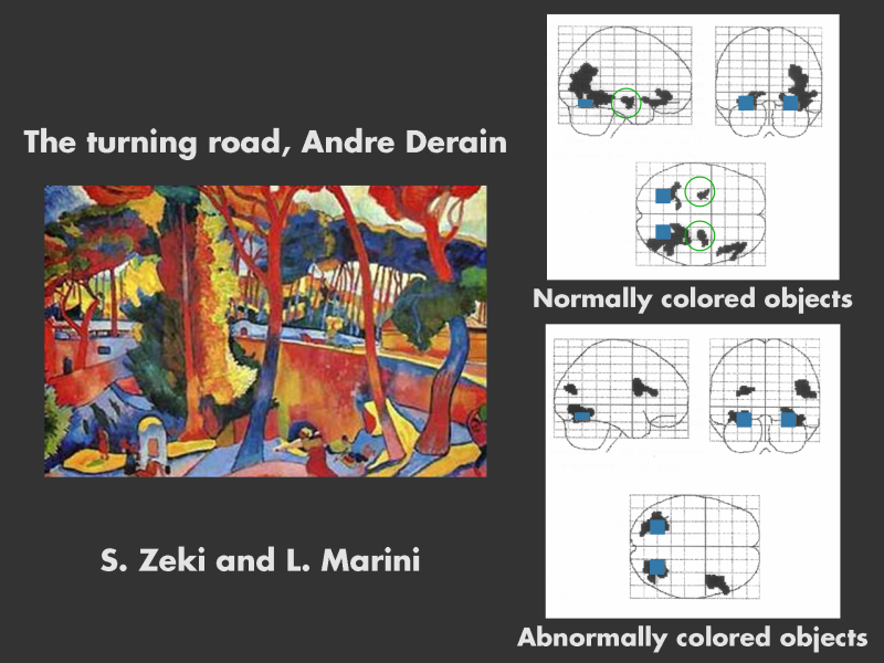
Here are the results of a clever recent experiment by Zeki and Marini . They were interested in visual pathways carrying color information. They showed subjects two scenes containing the same objects, one colored naturally and one colored abnormally, and then recorded their brain activity.
The top image on the right shows the brain activity when the subjects view a naturally colored scene. The visual areas V1 and V4 are active - these are marked with blue rectangles. In addition the hippocampus is activated ? it?s circled ; this area is implicated in memory. There is also activation in the frontal area of the right hemisphere.
Now repeat the experiment with the same objects, but abnormal colors. Now, there is no activity in the hippocampus, and the frontal activity is in a different region (middle frontal convolution). This frontal region is sometimes called the monitoring system.
The explanation is that the image with the normal colors is being recognized; whereas the one with abnormal colors is triggering a search and orienting response as we figure out what it is.
Zeki claims that artists are neurologists, experts in selectively activating visual pathways. The fauvist painters wanted to liberate color from shape and form; they appear to have done so successfully.